Our Science
Chemical Exchange Saturation Transfer Magnetic Resonance Imaging (CEST-MRI)
Magnetic resonance imaging (MRI) is a clinical diagnostic technique employing a variety of contrast mechanisms, some of which are reliant upon the mapping of differential water proton relaxation following radiofrequency excitation in a magnetic field. MRI has traditionally been applied to anatomical and functional imaging in the clinical setting, and has been moving toward molecular-level diagnosis in part through the use of biochemical-targeted contrast agents. Gadolinium-based chelates that reduce the relaxation time of neighboring water protons have previously been the focus of contrast agent development, however concerns about chelate stability in vivo has motivated a search for metal-free alternatives. Chemical Exchange Saturation Transfer (CEST) using organic (i.e. diamagnetic) probes is a recently proposed metal-free MRI technique offering a promising alternative mode of molecular-level contrast that can overcome the toxicity concerns associated with paramagnetic agents. In the Molecular Medicine Lab, we are exploring new chemistry enabling the development of conditional CEST contrast agents that only produce imaging signal when activated by biomolecular targets of interest.
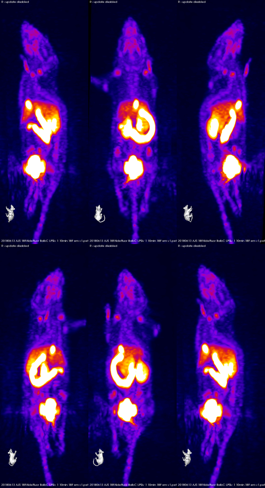
Positron Emission Tomography (PET) is another clinical diagnostic imaging technique demonstrating exquisite sensitivity. PET is reliant upon β decay of radioisotopes, releasing a positron that collides with nearby electrons to result in an annihilation event that is detected by the PET camera. Therefore, PET necessarily requires the injection of radioactive imaging agents (i.e. radiotracers) to provide for imaging signal. In the molecular medicine lab, we develop novel radiopharmaceuticals that expand the complement of endogenous biomarkers amenable to detection by PET. Our radiochemistry utilizes a range of isotopes, including 18F and 89Zr, to label small molecule and particulate radiotracers designed to image inflammation, metabolism, cancer, or immunomodulatory therapeutics.
Positron Emission Tomography (PET)
Fluorescent Fingerprinting Of Aldehydes As Point-Of-Care Diagnostics
Aldehydes can be innocuous contributors to normal biochemistry, however during disease or injury, a pathology-specific and more sinister complement of toxic aldehydes can form. This biochemical duality of biogenic carbonyls would make knowing the precise aldehyde present, i.e. whether it is friend or foe, in addition to total aldehyde concentration, important for the implementation of aldehydes as biomarkers of disease. The Molecular Medicine Lab is developing fluorogenic probes that rapidly complexes aldehydes in physiological solutions to provide an optical fingerprint in the form of excitation-emission matrices. Using data reduction and pattern-matching algorithms to analyse these fingerprints, the identification of the bound aldehyde biomarker from complex mixtures is possible. This unique combination of a fluorescent probe capable of determining both total aldehyde levels and identifying the bound species through a pattern matching routine lays the groundwork for novel implementations of chemometrics to low cost, rapid, and simply-implemented point-of-care diagnostics, which the Lab is actively pursuing.


